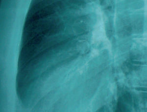
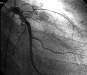
A 22-year-old man, occasional user of cocaine and smoker of 22 cigarettes/day, was referred to our center for chest pain. The physical examination and laboratory workup were normal. Electrocardiography showed right bundle branch block with Q-waves in V1, V2, and V3. The chest x-ray (Figure 1) revealed a round calcified image superimposed on the cardiac silhouette. On echocardiography, the left ventricle was found to be thinned and dilated, with general hypokinesia that was more evident in the septum and anterior aspect, and severe systolic dysfunction (ejection fraction, 30%). Coronary angiography showed complete occlusion of the left anterior descending artery distal to a giant calcified aneurysm (Figure 2) and complete occlusion of the right coronary artery. Dobutamine echocardiography demonstrated viable myocardium. The patient underwent a double aortocoronary bypass (left anterior descending artery to left mammary artery and posterior descending artery to right mammary). One year after surgery the patient's ejection fraction had improved (40%) and he had no symptoms of angina.

Figure 1.

Figure 2.
Although coronary aneurysms have been described in relation to cocaine use and in arteriosclerotic disease, the presence of a giant calcified aneurysm in this patient led to the presumptive diagnosis of prior Kawasaki disease.