Coronary artery aneurysm, with or without stenosis at the distal end, is a well-recognized characteristic of Kawasaki disease. The aneurysms show considerable variation in severity and progression.
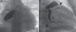
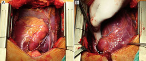
We present the imaging findings in a 10-year-old boy with an extreme case of this condition. The diagnosis of Kawasaki disease was established 2 years before the episode presented here. At that time, aneurysms had been detected in the proximal portion of the right (5 mm) and left (9 mm) coronary artery. Over time, the aneurysms had enlarged significantly (to 20mm and 23mm, respectively). Furthermore, the right coronary artery (RCA) became obstructed, the left anterior descending artery (LAD) developed severe stenosis, and the patient had an acute myocardial infarction. Angiography study showed a giant aneurysm and occlusion in the RCA, and a giant aneurysm and severe stenosis in the LAD. The circumflex artery (Cx) also demonstrated significant dilation, but without stenosis (Figure 1). In light of this situation, coronary surgery was performed (left internal mammary artery to LAD and right internal mammary artery to RCA). Intraoperative images show the RCA (Figure 2A) and the proximal portion of the LAD (Figure 2B). The postoperative progress was favorable and there were no adverse events.

