Sialadenitis is an extremely rare complication of procedures involving intravenous iodinated contrast media. To date, only 15 cases of iodide-associated sialadenitis have been described. These cases were reported in different clinical case scenarios, including a coronary angiogram (CA) procedure. The mechanism for iodide-induced sialadenitis may be either idiosyncratic or related to toxic accumulation of iodide in the salivary glands.
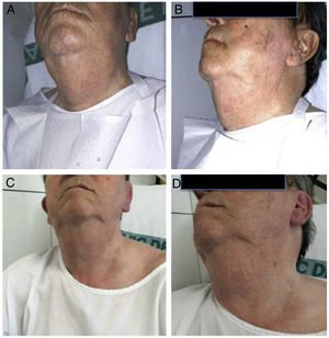
An 82-year-old man with previous aortic valve replacement and a saphenous vein graft to the circumflex coronary artery was referred to our center for a scheduled ambulatory CA to address progressive angina. Two previous CAs were performed without stenting, vascular, or anaphylactic complications. Six hours postprocedure, the patient noticed bilateral neck swelling predominantly on the right side. Physical examination revealed enlargement and a slight tenderness of the right submandibular gland (Figure A and B). Intravenous antihistamine and corticoids were initiated. A salivary gland ultrasound demonstrated homogenous inflammation of the right submandibular gland without dilated ducts and normal color velocity. These findings were compatible with noninfectious sialadenitis. The patient's clinical picture and echocardiographic abnormalities resolved after 3 days (Figure C and D).

Iodide sialadenitis is a rare late reaction to intravascular administration of iodine-containing contrast material leading to abnormal swelling of the salivary glands. The risk for this condition seems to be directly related to serum iodide levels; thus, renal insufficiency and a large iodide load are predisposing factors. Treatment of this condition is largely supportive, and most cases resolve in 2 to 4 days without treatment.
Given the widespread use of imaging and interventional techniques including CA that use iodinated contrast, cardiologists should be aware of this condition.