Publish in this journal
Journal Information
Vol. 74. Issue 8.
Pages 708 (August 2021)
Vol. 74. Issue 8.
Pages 708 (August 2021)
Image in cardiology
Full text access
Latest developments in 3D echocardiography. A novel tissue transparency tool
Novedades en ecocardiografía 3D: una nueva herramienta de transparencia tisular
Visits
519
Marina Pascual Izcoa,b,
Corresponding author
marinapasiz@hotmail.com

Corresponding author:
, Covadonga Fernández-Golfín Lobána,b, José Luis Zamorano Gómeza,b
a Servicio de Cardiología, Hospital Universitario Ramón y Cajal, Madrid, Spain
b Centro de Investigación Biomédica en Red de Enfermedades Cardiovasculares (CIBERCV), Spain
This item has received
Article information
Full Text
Download PDF
Statistics
Figures (3)
Show moreShow less
Additional material (5)
Full Text

Despite advances in 3D echocardiography, the technique still has some inherent limitations. New tools such as transillumination give images a more realistic appearance. This tool now includes the option of modifying tissue transparency and integrating color Doppler.

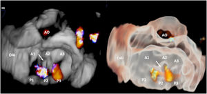
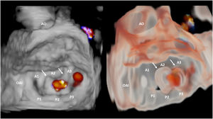
We present the case of a 75-year-old man with severe functional mitral regurgitation who underwent implantation of 2 mitral clip devices. The procedure was performed with 3D transesophageal echocardiographic guidance, using this new tissue transparency tool, which allowed identification (figure 1, video 1 of the supplementary data) of the first clip (white arrow) at A2-P2 in a somewhat medial position. With color Doppler, 2 residual regurgitation jets could be seen (figure 2, right; video 2 of the supplementary data). Note the difference in detail of valve and adjacent structure anatomy (LAA, left atrial appendage; Ao, aorta) between the tissue transparency tool (figure 2, right; video 2 of the supplementary data) and the conventional 3D reconstruction (figure 2, left; video 3 of the supplementary data). In fact, after the second clip was implanted, standard 3D reconstruction did not allow adequate identification of the devices (figure 3, left; video 4 of the supplementary data). This case illustrates how this new tissue transparency tool makes it possible (figure 3, right; video 5 of the supplementary data), for the first time, to identify structures that are difficult to visualize (mitral clips) even with other modern 3D-echo techniques

Figure 1
(0.11MB).
Figure 2
(0.18MB).
Figure 3
(0.21MB).
Acknowledgements

The authors thank Drs A. Sánchez-Recalde and L. Salido Tahoces for their collaboration.

Appendix A
APPENDIX. SUPPLEMENTARY DATA

Supplementary data associated with this article can be found in the online version available at https://doi.org/10.1016/j.rec.2020.11.016

Appendix B
SUPPLEMENTARY MATERIAL

(0.5MB)
(0.49MB)
(0.46MB)
(0.29MB)
(0.44MB)

Copyright © 2020. Sociedad Española de Cardiología
Idiomas
Revista Española de Cardiología (English Edition)

Subscribe to our newsletter

View newsletter history
Article options
Tools
Supplemental materials
es en

¿Es usted profesional sanitario apto para prescribir o dispensar medicamentos?

Are you a health professional able to prescribe or dispense drugs?