The effectiveness of intraventricular synchrony correction by cardiac resynchronization therapy (CRT) has been correlated with electrocatheter (EKT) positioning in the coronary veins. Multidetector computed tomography (MDCT) with contrast administration allows precise, noninvasive visualization of the coronary arteries, as well as the anatomy and distribution of the coronary veins. With this technique it is possible to identify the most suitable vein and provide the electrophysiologist with a guide for implanting the left ventricular EKT.
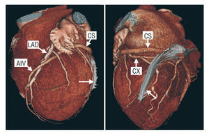
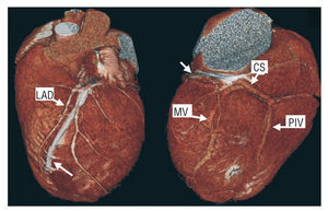
The common causes of failure of this therapy include absence of a suitable coronary vein and errors in EKT implantation. Current data indicate that an optimal clinical and hemodynamic response is obtained in most patients when the left ventricular EKT is placed in a lateral or posterolateral position. This was the case of one of our patients, who underwent MDCT with a 64-detector Toshiba Aquilion scanner and had a good response to CRT. The catheter was visualized in the marginal vein (arrow) in a three-dimensional reconstruction of the heart, analyzed from 2 angles (Figure 1). As is observed in Figure 2, incorrect positioning of the EKT (arrow) can sometimes be detected in MDCT images, in this case in the anterior interventricular vein, which runs parallel to the left anterior descending artery (LAD), despite the presence of an adequate marginal vein (MV). This fact would explain the inappropriate response to resynchronization therapy experienced by this patient.
Figure 1. Cx indicates circumflex artery; LAD, left anterior descending artery; CS, coronary sinus; AIV, anterior interventricular vein (great cardiac vein); PIV, posterior interventricular vein (middle cardiac vein).
Figure 2. LAD indicates left anterior descending artery; CS, coronary sinus; MV, marginal vein; PIV, posterior interventricular vein (middle cardiac vein).


