
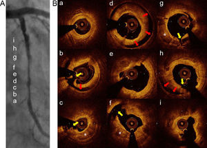
A 58-year-old man presented with stable angina pectoris. Coronary computed tomography angiography and coronary angiography revealed significant diffuse stenosis suggestive of coronary artery calcification at the proximal to midleft circumflex artery (Figures 1A, 1B). Percutaneous coronary intervention was performed. Frequency-domain optical coherence tomography revealed calcified plaques (Figure 1C, asterisk). At the distal-to-mid portion of the culprit lesion, moderate calcified plaques (calcium arc < 180°) were observed (a, b and c). At the mid-to-proximal portion, severe calcified plaques (calcium arc ≥ 180°) were observed (d and e). Then, balloon dilatation using a 2.5mm×10mm Scoreflex scoring balloon catheter (OrbusNeich, Hong Kong, China) was performed. Coronary angiography after scoring balloon dilatation demonstrated diffuse coronary artery dissection with a radiolucent flap (Figure 2A). Frequency-domain optical coherence tomography nicely depicted the entry tear, intimal-medial flap, and false lumen reaching to the media (Figure 2B). Yellow arrows show the intimal-medial tear producing a communication between the true and the false lumen. Red arrows show the dissection reaching the media. Two stents were placed and final TIMI flow grade 3 was achieved.


In this case, the extent of vessel calcification and therapeutic dissection after scoring balloon dilatation were observed by frequency-domain optical coherence tomography. This procedure allows more accurate assessment of the extent of calcified plaque close to the lumen than intravascular ultrasound because calcified plaque observed by intravascular ultrasound causes acoustic shadowing. Thus, we think that for percutaneous coronary intervention in calcified lesions, optical coherence tomography can be used to assess calcified plaque and the effect of new technologies such as the scoring balloon.