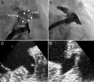
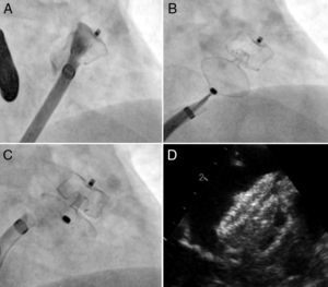
Percutaneous closure of the left atrial appendage (LAA) is becoming much more widespread in Spain. Although the LAA varies greatly in its morphology, 4 anatomical subgroups have been proposed: a) windsock; b) cauliflower; c) cactus, and d) chicken wing. In most cases, LAA morphology does not determine the implantation technique used, as the main anatomical variable is its distality. The case presented shows an LAA whose morphology did not fall into any of the aforementioned categories and whose closure required recourse to nonstandard techniques. The anatomy was characterized by a short (< 15mm) and narrow landing zone, with 2 small, symmetric lobes opposite one another (Figure 1). Bearing in mind that the target area had a diameter of 13 to 14mm and a depth of 11mm (Figure 1), the device chosen for implantation was a 16-mm Amplatzer Amulet. After formation of the initial triangle (Figure 2A), we attempted to enlarge the target zone by pushing gently on the delivery sheath at the same time as the device's lobe was deployed. A characteristic of the Amulet device is that it allows the LAA walls to be displaced forward with a low risk of perforation as, once the triangular form is adopted, the distal pin is invaginated at the same time as the lobe is deployed (Figure 2B). After the stability and position of the device was checked, it was released with no complications (Figure 2C and Figure 2D).
CONFLICTS OF INTEREST

X. Freixa is a proctor for St. Jude Medical.