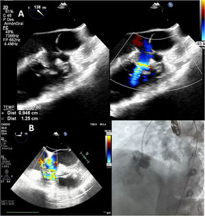

We present the case of a 15-year-old male patient with a restrictive subaortic ventricular septal defect (VSD) and a large aneurysm protruding into the right ventricle that caused moderate tricuspid regurgitation (TR) (figure 1, video 1 of the supplementary data). In line with the latest guidelines, percutaneous treatment was selected, given the favorable anatomy (distance to aortic leaflets > 1cm).

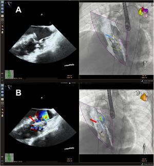
The procedure was performed under general anesthesia and was guided by transesophageal echocardiography (EPIQ system, Philips Healthcare, The Netherlands). Because the defect was 12.5mm in size (figure 1A), a 14-mm KONAR-MF device (Lifetech, China) was applied with a retrograde approach. Catheterization of the VSD was complicated by the size of the aneurysm. Accordingly, the EchoNavigator fusing imaging system was used. The echocardiographic projection comprised a 3-chamber view at 122° with left anterior oblique fluoroscopic projection at 42° and cranial at 18°. This obtained a good overlapping echocardiographic and fluoroscopic view of the VSD, which facilitated rapid catheterization (figure 2A; the blue arrow indicates the guidewire path; this figure is shown in color only in the online version of the article). Next, the guide catheter was advanced (figure 2B, the red arrow indicates the catheter path; video 2 of the supplementary data) and the device was deployed. All maneuvers were monitored via fusion imaging (video 3 of the supplementary data).

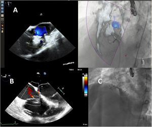
The final outcome was excellent, without a residual shunt or complications and with an almost entirely resolved TR (figure 3, video 4 of the supplementary data). As far as we know, this is the first reported case of complex VSD closure with the KONAR-MF device and exclusively guided by fusion imaging.

The patient's consent was obtained for dissemination of the case.
FundingThe present work has not received funding.
Authors’ ContributionsI.-A. Nistor contributed to the conception, data acquisition, manuscript drafting, and revision and assumes responsibility for all aspects of the article and the veracity of all parts of the work. D. Mesa Rubio and M. Pan Álvarez-Ossorio contributed to the conception, design, and critical revision of the intellectual content and assume responsibility for all aspects of the article and the veracity of all parts of the work.
Conflicts Of InterestThere are no conflicts of interest.
Supplementary data associated with this article can be found in the online version, at https://doi.org/10.1016/j.rec.2023.07.017