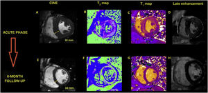
We describe the case of a 25-year-old man admitted to hospital due to spontaneous subarachnoid hemorrhage that rapidly progressed to cardiogenic shock. The patient had elevated cardiac enzymes and depressed ST segment in the lower and precordial leads. Emergency echocardiography showed a nondilatated left ventricle with severe systolic dysfunction (left ventricular ejection fraction [LVEF], 15%) secondary to general hypokinesia that was more pronounced in the basal segments. Consequently, cardiomyopathy was suspected due to inverted stress. The patient's clinical progress was favorable during the first week, with LVEF recovery. The study was completed with cardiac magnetic resonance imaging on hospitalization day 10, revealing severe concentric hypertrophy in the cine sequences (figure 1A), with normal LVEF despite severely impaired longitudinal contraction. Parametric maps showed diffusely elevated T1- and T2-weighted values (figure 1B: T2, 56-57ms; normal value [NV], <50ms; figure 1C: T1, 1402ms; NV, 1207±54ms) and a high extracellular volume (ECV) fraction (35%; NV, <30%). The images also showed diffuse and very faint intramyocardial late gadolinium enhancement (figure 1D). These findings indicated generalized myocardial edema consistent with stress cardiomyopathy; however, acute myocarditis and underlying hypertrophic cardiomyopathy were also considered. Surprisingly, 6-month follow-up cardiac magnetic resonance showed complete regression of the hypertrophy (figure 1E), normalization of the T2- (figure 1F: T2, 44-46ms) and T1-weighted segments (figure 1G: T1, 1207ms) as well as of extracellular volume (28%), plus complete absence of late gadolinium enhancement (figure 1H). In conclusion, serial cardiac magnetic resonance imaging can greatly aid the differential diagnosis of stress cardiomyopathy, which may be confused with other similar clinical entities.

The patient gave consent for publication of his case study and scientific dissemination of the cardiac MRI images.
FUNDINGNone.
AUTHORS’ CONTRIBUTIONSAll authors contributed equally to the discussion and text of this case study.
CONFLICTS OF INTERESTNone.