A 69-year-old man was admitted for fever 2 weeks after implantation of an automatic defibrillator for primary prevention of ischemic cardiomyopathy. He had a hematoma and inflammation at the generator pocket, and acute-phase reactants were elevated. Transesophageal echocardiography ruled out valvular and device lead vegetations. Cultures of blood and the hematoma material were negative. Nonetheless, the fever persisted, and to complement the diagnosis we performed 18F-FDG positron emission tomography combined with cardiac computed tomography angiography (PET/CT angiography).
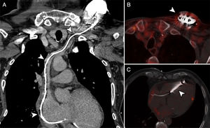
Prospective electrocardiogram (ECG)-triggered cardiac CT angiography showed no vegetations/thrombi at the lead (Figure 1A, arrows). Anatomic-metabolic fusion images excluded pathologic uptake at the generator and lead (Figure 1B and 1C, arrows), whereas mild, homogeneous hypermetabolism was seen around the generator (Figure 1B, arrow), attributable to the recent postoperative changes.

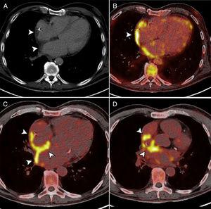
CT angiography also showed wall thickening of both atria and reticulation of the interatrial fat (Figure 2A, arrows). The fusion images depicted marked hypermetabolism of the atrial walls, interatrial septum, pericardium, and tissue surrounding the superior vena cava, together with signs of pericarditis (pericardial effusion with focal 18F-FDG uptake) (Figure 2B-2D, arrows). The final diagnosis was inflammatory response syndrome following device implantation, with pericarditis, and atrial wall and perivascular involvement. Anti-inflammatory treatment was started, the condition resolved, and device infection was ruled out.

This case illustrates the usefulness of PET/CT angiography to confirm or exclude inconclusive cases of intracardiac device infection and provide alternative diagnoses, such as postimplantation inflammatory response syndrome.