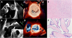

A 76-year-old woman presented with a recent diagnosis of atrial fibrillation. A transthoracic echocardiogram was performed, excluding relevant structural heart disease beyond mild biatrial enlargement, but a large mass (33 x 25mm) was observed in the left atrium. For further characterization, a transesophageal echocardiogram was ordered, confirming the presence of a large, round, smooth mass, with heterogeneous density, arising from the posterior mitral leaflet, suggesting a myxoma with atypical location (figure 1A-C, ). However, routine examination of the aortic valve revealed small mobile structures (figure 1B, ). Visualization with standard 3-dimensional imaging provided no additional information, but further processing of the image with transillumination echo allowed us to clearly identify the presence of several filiform structures anchored in the left coronary leaflet (maximum length 9 mm), suggesting multiple fibroelastomas (figure 1D, ). The patient consented to the publication of these images.

The patient underwent subsequent surgery for removal of these masses. In addition to the atriotomy, an arteriotomy was performed and examination of the aortic valve revealed several fibrillar structures on its aortic face. The pathology analysis confirmed the presence of both cardiac myxoma (figure 1E) and papillary fibroelastoma (figure 1F).
Transillumination is a new technology that improves the quality of 3-dimensional images and can provide better diagnostic accuracy. A complete examination of all cardiac structures is desirable beyond the structure of interest, especially when cardiac surgery is to be performed. This case highlights how a thorough evaluation is important to detect rare or unexpected findings, such as, in this patient, 2 tumors in 1 heart.
FUNDINGNo funding was received.
AUTHORS’ CONTRIBUTIONSS. Valbuena-López and V. Juárez Olmos obtained and processed the images and drafted the text. E. Pena Burgos contributed to the production of the figure and reviewed the manuscript.
CONFLICTS OF INTERESTNone to disclose.
Supplementary data associated with this article can be found in the online version, at https://doi.org/10.1016/j.rec.2022.01.005