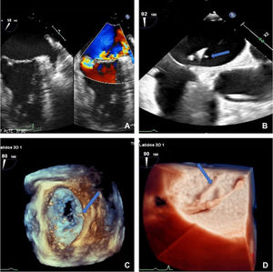
We present the case of an 83-year-old woman with chronic heart failure in New York Heart Association functional class II-III secondary to severe primary mitral regurgitation due to P3 rupture, with no other relevant history (figure 1A). Given the high surgical risk, the multidisciplinary team decided on percutaneous treatment of the mitral regurgitation.

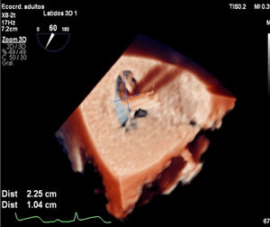
After transseptal puncture, on transesophageal echocardiography, an image was observed adhered to the MitraClip guide catheter, which was at first suspected to be a thrombus (blue arrow) (figure 1B and ). On 3D echocardiography (figure 1C: with aorta orientated at 9 o’clock for better visualization) and images with transillumination (figure 1D), the image was observed to be dependent from the interatrial septum, raising the need for a differential diagnosis (). Transillumination is a rendering of 3D echocardiography in which, using a movable virtual light position, the detail and depth perception of an image can be improved. In this case, it provided better visualization of the tear and of a complex iatrogenic interatrial communication, buttonhole-shaped and measuring 10×22mm, with a significant left-right shunt (figure 2).

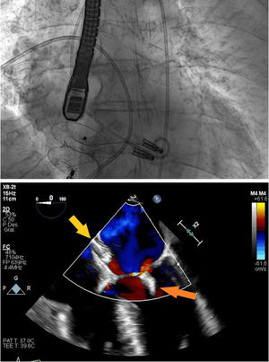
We proceeded to implant 2 MitraClip devices, with a good final result, minimal mitral regurgitation, and no significant gradients. Finally, the interatrial communication was corrected with a 28-mm Amplatzer device, the filamentous septal remains were captured and there was no final residual shunt either on angiography or echocardiography (figure 3; yellow arrow, Amplatzer; orange arrow, MitraClip).

Supplementary data associated with this article can be found in the online version available at https://doi.org/10.1016/j.rec.2020.08.024