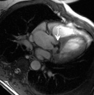
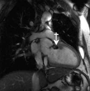
A 46-year-old patient who consulted for palpitations was found to have isolated repeated premature ventricular beats on the electrocardiogram. Transthoracic electrocardiography showed a subaortic membrane that did not generate a gradient in that area, as well as a slightly dilated ascending aorta (maximum diameter, 44 mm). To complete the study, cardiac magnetic resonance imaging was performed with dynamic gradient echo sequences (fast-imaging employing steady-state excitation [FIESTA] technique). A hypointense image consistent with a fibrocalcific mass was detected in the left ventricular outflow tract (LVOT), which ran in a subaortic course to the valve plane and continued along the intramyocardial interventricular septum. The patient had a tricuspid aortic valve that was functionally bicuspid due to calcification of the commissure between the left and right coronary leaflets. The 5-chamber view at the level of the LVOT (Figure 1) shows the subaortic membrane (arrow) continuing to the valve plane. The extension of the fibrocalcification along the long axis of the interventricular septum (arrow) is seen in Figure 2. Because there were no relevant symptoms and no gradient indicating hemodynamic compromise, these imaging findings led us to take a conservative approach consisting in imaging surveillance every 6 months to monitor the evolution of the condition.

Figure 1.

Figure 2.