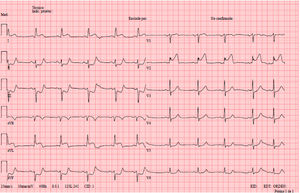

An 80-year-old man with diabetes, hypertension, and dyslipidemia and a former smoker attended the emergency department with central thoracic pain with typical characteristics. Upon his arrival at our center, an electrocardiogram was performed (figure 1), which revealed ST-segment elevation in leads I, aVL, and V2 with decreases in lower leads. He was therefore transferred to the catheterization laboratory for primary angioplasty.

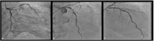
During the procedure, 2-vessel coronary artery disease was seen (figure 2A): the circumflex artery had a chronic occlusion and the anterior descending (AD) artery had a third-degree lesion with distal flow (TIMI 2) together with an occluded first diagonal branch (TIMI 0). Both lesions were treated (figure 2B,C), with a drug-eluting stent implanted in the AD artery and angioplasty performed with a drug-eluting balloon in the diagonal branch. The patient was discharged without symptoms 3 days afterward.

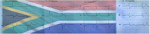
The interest of this case lies in the presence of a rare sign: “the South Africa flag sign” (figure 3, with the flag superimposed on the first electrocardiogram performed in the health center). It is associated with high lateral infarction, and the artery typically involved is the first diagonal branch, which, because it passes by the anterolateral face of the left ventricle, is characterized by ST-segment elevation in leads I, aVL, and V2 and a decrease in lead III. Knowledge and recognition of this particular tracing is important because, due to the lack of ST-segment elevation in contiguous leads, it can be missed, with fatal consequences.

The patient was informed and gave his consent for publication of the case.
FUNDINGNone.
AUTHORS’ CONTRIBUTIONSAll authors participated in the design, drafting, and revision of the final manuscript.
CONFLICTS OF INTERESTThe authors have no conflicts of interest in relation to this manuscript.