
A 72-year-old man with a diagnosis of sinus node dysfunction and no other known cardiac anomalies was referred for implantation of a dual-chamber pacemaker. The implant was performed through a right subclavian approach. During the procedure the guidewires were seen to cross in the contralateral side (Figure 1) and run along the course of the coronary sinus, which led to the diagnosis of persistent left superior vena cava, with atresia of the right vena cava. For this reason, the electrodes were placed in a retrograde fashion through the coronary sinus into the right chambers (Figure 2). The ventricular electrode was left in the right ventricle, preshaping the guidewire in a loop and without any fixation, whereas active fixation was used for the atrial electrode. Later, the thresholds and functioning of the system were checked and found to be correct.

Figure 1.

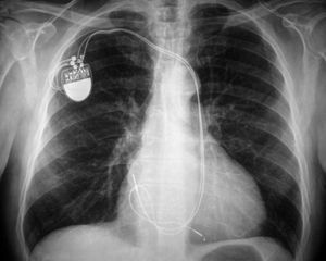
Figure 2.
Persistent left superior vena cava is a congenital anomaly that occurs in 0.1%-0.3% of individuals with a normal heart and in approximately 3% of patients with congenital heart disease. The anomaly has no clinical relevance, although electrode placement may be more complex when there is associated superior vena cava atresia.