A sixty-one-year-old man with a history of arterial hypertension went to the emergency room secondary to intense interscapular pain, suggestive of dissection. Physical examination revealed diaphoresis, a systolic arterial pressure of 165 mm Hg, and symmetric peripheral pulses.
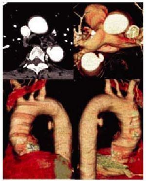
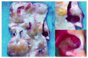
Electrocardiogram (ECG) revealed a sinus rhythm with signs of left ventricular hypertrophy. Thoracic computerized tomography (CT) with 3-dimensional reconstruction of the large vessels showed an adventitial hematoma of the proximal third of the descending thoracic aorta with a 16×10 mm sacular penetrating ulcer on the anterior face. Imaging of an endoluminal dissection showed the diameter of the thoracic aorta to be 33 mm (Figure 1). During surgery external examination revealed a thoracic aorta with a subadventitial hematoma in the upper half; resection of approximately 6 cm of the aortic portion was performed and replacement was made with a Dacron prosthesis. The excised portion revealed an atherosclerotic aorta with a hematoma on the wall and an opening on the endothelial wall corresponding to a sacular pseudoaneurysm. This was associated with multiple ulcerations on the remainder of the aortic wall (Figure 2).
Fig. 1
Fig. 2.


