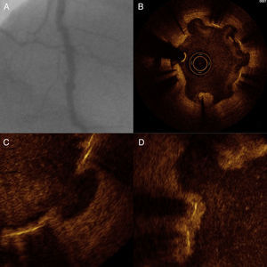
A 41-year-old male smoker with hypertension underwent diagnostic angiography for angina on exertion. The study showed chronic occlusion of the proximal left anterior descending artery (LAD) with collateral circulation from the right coronary artery. Stress echocardiography showed viability and inducible ischemia in the affected territory. Angioplasty was performed, with implantation of 4 sirolimus-eluting stents (SES) of 2.25×23, 2.25×28, 2.5×13, and 2.5×23mm, with angiographic success. Eighteen months later, the patient was readmitted for exertional angina, and coronary angiography was again performed. The LAD was patent, but had an irregular appearance with slow clearance along the length of the treated segment (Fig. 1A). Optical coherence tomography (Fig. 1B) showed vascular wall retraction and an ulcerated appearance around the stent. The struts were not affected and, for the most part, showed re-endothelization (Fig. 1C and D). The vessel lumen showed no significant stenosis.

Contrast retention on angiography following implantation of a drug-eluting stent has been associated with intracoronary imaging findings of late stent malapposition. The vascular wall reaction to SES has been described as “peri-stent ulcer like appearance” (PSUA). It has a reported incidence of up to 50% in small series and is speculated to be a harbinger of late malapposition.