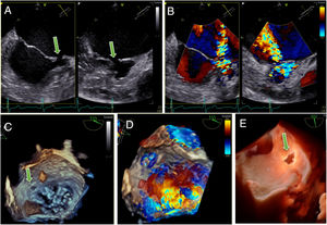

We present the case of a 52-year-old patient with lupus and chronic leishmaniosis diagnosed with symptomatic mitral regurgitation at another center. The patient was referred to evaluate percutaneous edge-to-edge mitral valve repair in our center due to comorbidities. Transesophageal echocardiography showed normal function of the mitral valve and the presence of a left ventricle to left atrium shunt located next to the antero-lateral annulus (figure 1, ). This generated an eccentric jet that swept along the mitral coaptation line mimicking severe mitral regurgitation in transthoracic echocardiography (). The etiology of the shunt was not clear, but we hypothesized that it could be secondary to a previous drained paravalvular abscess since the patient had a long history of immunosuppressive treatment and several episodes of fever in the setting of chronic leishmaniosis.

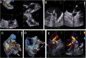
The patient was presented at a heart team session, and it was decided to treat the shunt percutaneously with a plug. The intervention (figure 2) was conducted following the steps of a paravalvular prosthetic mitral paravalvular leak closure. Echo-guided transseptal access was performed to introduce a deflectable catheter in the left atrium, which was guided toward the atrial orifice of the fistula (figure 1, arrows) to cross the defect (). After 3-dimensional echo measurements, an Amplatzer Vascular Plug III 14 x 5 mm (Abbott, United States) (figure 2 and figure 3, arrows) was successfully implanted with a mild-to-moderate residual shunt across the device (). The 3-month follow-up transesophageal echocardiography (figure 3, ) showed significant improvement with only minimal residual shunt. Informed consent was obtained by telephone.
FUNDING

No funding to disclosure.
AUTHORS’ CONTRIBUTIONSL. Sanchis performed acquisition of images and elaborated the figures. L. Sanchis, B. Vidal and X. Freixa wrote and reviewed the text.
CONFLICTS OF INTERESTL. Sanchis and X. Freixa are proctors of Abbott for Mitraclip/Triclip implantation (Abbott, United States). L. Sanchis is associate editor of Rev Esp Cardiol. The journal's editorial procedure to ensure impartial handling of the manuscript has been followed.
Supplementary data associated with this article can be found in the online version available at https://doi.org/10.1016/j.rec.2021.11.010