

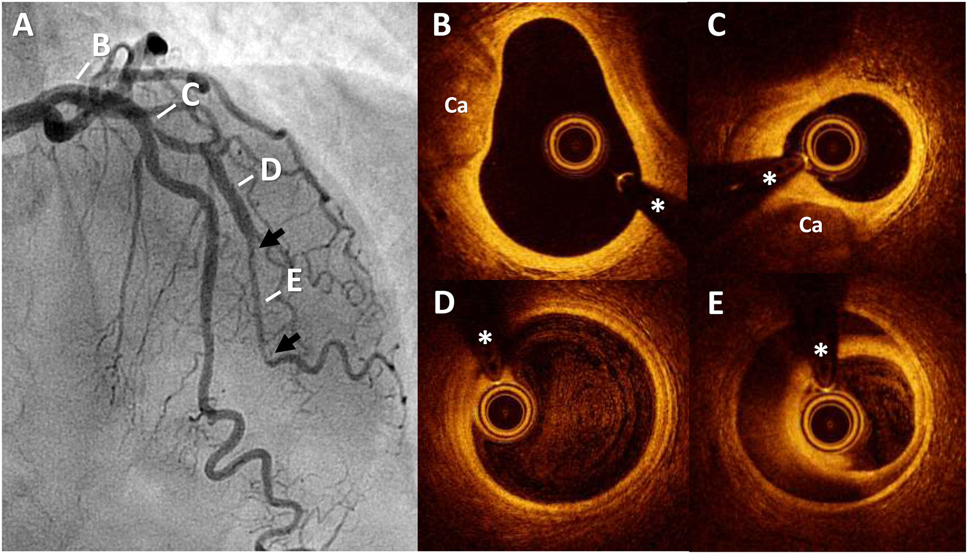
A 63-year-old woman with hypertension and an active smoker was admitted to our hospital with non–ST-segment elevation acute myocardial infarction. Emergency coronary angiography (figure 1A) revealed tortuous arteries, with a calcified lesion at the ostium of the first diagonal branch and, more distally, an abrupt loss of caliber and straightening of the vessel with distal caliber recovery (arrows). Optical coherence tomography showed calcified atherosclerosis (figure 1B; Ca) in the proximal anterior descending artery and at the origin of the diagonal branch (figure 1C). After a completely healthy arterial segment (figure 1D), a characteristic intramural hematoma was evident (figure 1E; asterisks indicate guidewire artifacts). The patient was managed conservatively and showed an excellent clinical response to 12-month dual antiplatelet therapy without beta-blockers. Dissection within an atherosclerotic plaque is excluded from the definition of spontaneous coronary artery dissection (SCAD), given its distinct etiology and management. However, as this case illustrates, patients can simultaneously present both stable atherosclerotic disease and SCAD in healthy arterial segments. In this context, intracoronary imaging is essential for establishing the differential diagnosis. There have been no other documented cases of intracoronary imaging of SCAD in arteries that also show significant atherosclerosis in other segments. In this case, a concordant epidemiological scenario and high diagnostic suspicion aided the presumptive diagnosis but, in arteries with atherosclerosis, only intracoronary imaging offers certain diagnosis of this surprising clinical entity.
FUNDING
None.
ETHICAL CONSIDERATIONSInformed consent was obtained from the patient for the complementary tests performed and for this publication. Involvement of an ethics committee was not necessary. Possible sex and gender biases have been taken into account.
STATEMENT ON THE USE OF ARTIFICIAL INTELLIGENCEArtificial intelligence has not been used in the preparation of this work.
AUTHORS’ CONTRIBUTIONSAll authors have contributed to the drafting, conception, and revision of the manuscript.
CONFLICTS OF INTERESTNone.